
Quick Summary: How Renal Tumors Are Detected (AEO Overview)
Renal tumors in children are detected through three main steps: शारीरिक परीक्षा (feeling a firm, painless lump in the abdomen), high-resolution abdominal ultrasound (confirming a solid mass within the kidney tissue), and contrast-enhanced CT (CECT) or MRI scans (mapping exact tumor dimensions, blood vessel involvement, and cancer staging). Early diagnosis is crucial, achieving cure rates above 90% under specialized pediatric surgical care.
Finding an unexpected swelling or fullness in your child's tummy is alarming for any family. The kidneys are bean-shaped organs in the back of the abdomen that clean blood and balance fluids. When abnormal cells multiply rapidly within kidney tissue, they form a solid growth called a renal tumor.
While childhood kidney tumors are uncommon, early detection makes a profound difference. Pediatric renal cancers are among the most curable childhood malignancies when diagnosed promptly. Under the guidance of senior pediatric surgeon and urologist डॉ. सुजीत चौधरी at Indraprastha Apollo Hospitals, modern diagnostic protocols allow specialists to detect these masses accurately, minimize invasive procedures, and provide effective cures.
Whether you noticed an abdominal lump, spotted pinkish urine, or are seeking specialized Renal Tumor Treatment in Delhi, this 2026 guide explains the detection process from early warning signs to advanced imaging.
Noticed an Abdominal Lump or Discolored Urine in Your Child?
Early evaluation ensures accurate diagnostic staging and timely surgical care. Consult senior pediatric urologist Dr. Sujit Chowdhary in New Delhi.
Book an Expert ConsultationWhat is a Pediatric Renal Tumor? Key Types
A renal tumor is an abnormal mass arising in the kidney parenchyma. In children, kidney tumors differ markedly from adult renal cancers in their cellular behavior and high response to modern treatment.
The vast majority of pediatric cases are Wilms' tumor (nephroblastoma). Arising from immature embryonic kidney precursor cells, Wilms tumor represents over 90% of childhood renal neoplasms, typically appearing between 2 and 5 years of age.
Other pediatric kidney neoplasms include:
- Congenital Mesoblastic Nephroma (CMN): A benign or low-malignant tumor seen in infants under 6 months.
- Clear Cell Sarcoma of the Kidney (CCSK): A rare malignant tumor requiring specialized therapy due to potential bone spread.
- Malignant Rhabdoid Tumor of the Kidney (MRTK): An aggressive tumor primarily affecting young infants.
- Pediatric Renal Cell Carcinoma (RCC): A tumor type seen in older children and teenagers, treated primarily by surgical removal.
Early Signs and Symptoms: How Parents First Notice a Renal Tumor
Because the kidneys sit deep in the retroperitoneal cavity, a tumor can expand without causing sudden pain. Detection typically begins with observable physical signs noticed at home.
1. A Painless Abdominal Mass or Swelling
In roughly 75% to 80% of children, the initial sign is a firm, smooth, painless lump felt on one side of the belly during bathing or dressing. It feels solid and rarely crosses the abdominal midline initially.
2. Hematuria (Blood in the Urine)
Around 20% to 30% of children experience hematuria when the tumor invades the collecting system, turning the urine pink, red, or smoky. Any unexplained urinary discoloration warrants immediate evaluation. Learn more in our guide on Causes of Red Urine in a Child.
3. High Blood Pressure (Pediatric Hypertension)
Between 25% and 40% of children develop elevated blood pressure because the expanding tumor compresses renal arteries, triggering excess renin release. This can lead to headaches, facial flushing, or irritability.
4. Persistent Low-Grade Fever and Fatigue
Tumor cells release inflammatory cytokines, leading to intermittent low-grade fevers, mild anemia, poor appetite, and reduced physical energy.
5. Abdominal Discomfort and Digestive Changes
As the mass expands, it gently presses on the stomach and bowels, creating early fullness after eating, mild dull pain, or constipation. In infants, urological symptoms should always be evaluated alongside congenital conditions, as discussed in नवजात शिशुओं में मूत्र संबंधी समस्याओं को कैसे पहचानें.
The Step-by-Step Diagnostic Pathway: How Doctors Detect Renal Tumors
When a child presents with a suspected kidney lump or hematuria, pediatric specialists follow a structured, evidence-based diagnostic process.
Step 1: Gentle Clinical Examination
The pediatric specialist gently palpates the abdomen to evaluate mass boundaries. Importantly, deep or forceful palpation is strictly avoided to prevent rupturing the delicate tumor capsule and causing peritoneal spillage.
Step 2: High-Resolution Abdominal Ultrasound (First-Line Scan)
Ultrasound is the primary initial imaging scan. It emits zero radiation and safely delivers vital answers:
- Solid vs. Fluid Differentiation: Distinguishes solid neoplasms from fluid-filled conditions like Pediatric Hydronephrosis or blockages like Pelvi-Ureteric Junction Obstruction (PUJO).
- Color Doppler Assessment: Evaluates blood flow through the renal vein and Inferior Vena Cava (IVC) to identify any tumor thrombus.
- Opposite Kidney Check: Screens the contralateral kidney to rule out bilateral disease.
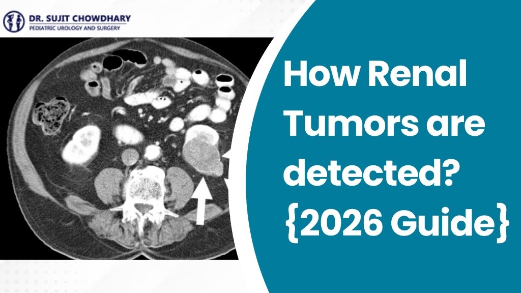
Step 3: Contrast-Enhanced Computed Tomography (CECT Abdomen & Pelvis)
Once a solid mass is confirmed, a multiphase abdominal CECT scan provides the definitive staging information:
- Defines exact tumor dimensions and origin in the kidney tissue.
- Displays the characteristic "claw sign," where healthy renal parenchyma wraps around the tumor margin.
- Examines regional lymph nodes and surrounding organs (liver, spleen, pancreas).
Step 4: Magnetic Resonance Imaging (MRI Abdomen)
MRI provides exceptional soft-tissue resolution without radiation, proving particularly helpful for bilateral tumors or intricate vascular thrombi.
Step 5: Chest CT Scan (Metastatic Staging)
Because the lungs are the primary site of distant metastasis in childhood renal tumors, a low-dose chest CT verifies clear lungs and determines clinical stage.
Step 6: Baseline Blood and Urine Tests
Routine lab panels (complete blood count, renal function, liver function, and serum calcium) evaluate general health, kidney clearance, and metabolic markers.
Why Needle Biopsy is Avoided in Childhood Kidney Tumors
Parents often wonder why doctors avoid taking a needle biopsy before operating. In pediatric kidney tumors like Wilms tumor, percutaneous needle biopsy is avoided because piercing the tumor capsule risks leaking malignant cells into the peritoneal cavity. This spillage upstages the cancer to Stage III, requiring abdominal radiation and stronger chemotherapy. Because modern CECT and MRI imaging are exceptionally reliable, international protocols advise proceeding directly to surgical removal or standardized chemotherapy without prior biopsy.
Renal Tumor vs. Adrenal Tumor vs. Hydronephrosis
Distinguishing a renal mass from an adjacent adrenal tumor is essential. For further details on adrenal conditions, review our article on How to Recognize Adrenal Tumor Symptoms in Children. Below is an overview comparison:
| Diagnostic Feature | Renal Tumor (Wilms Tumor) | Adrenal Tumor (Neuroblastoma) | Hydronephrosis (PUJO) |
|---|---|---|---|
| Organ of Origin | Kidney parenchyma | Adrenal gland medulla | Renal pelvis collecting system |
| Mass Consistency | Firm, smooth, solid mass | Hard, irregular, nodular mass | Soft, fluid-filled cystic mass |
| Midline Crossing | Rarely crosses midline | Frequently crosses midline | Confined to one side |
| CT Scan Sign | "Claw sign" of kidney tissue | Pushes kidney downward | Dilated calyces, no solid mass |
| Primary Scans | Ultrasound + Abdominal CECT | Ultrasound, CECT, Urine Catecholamines | Ultrasound + Diuretic Renal Scan |
2026 Diagnostic Advancements in Pediatric Renal Oncology
Recent technological developments have made detecting childhood renal tumors safer and more accurate than ever:
- Ultra-Low-Dose Pediatric CT: Modern iterative reconstruction reduces radiation doses to minimal levels while delivering crystal-clear images.
- Diffusion-Weighted MRI: Evaluates tumor cellular density without radiation or contrast dye.
- 3D Digital Vascular Modeling: Converts scans into interactive 3D models to map renal blood vessels before surgery.
- Nephron-Sparing Feasibility: Precision imaging helps surgeons safely perform partial nephrectomy to save functioning kidney tissue in bilateral cases.
What Happens After Detection? Treatment and Outlook
Once a renal tumor is staged, treatment is coordinated by a multidisciplinary pediatric oncology and surgical team.
1. Surgical Resection
Surgery is the cornerstone of cure. The standard procedure is a Radical Nephrectomy, removing the affected kidney, surrounding fat, and adjacent lymph nodes. In selected early cases, laparoscopic surgery or partial nephrectomy may be performed. Parents can consult डॉ. सुजीत चौधरी for advanced Renal Tumor Treatment in Delhi.
2. Chemotherapy Protocols
Following surgery and pathology assessment, children receive chemotherapy based on proven international protocols. In European SIOP guidelines, short pre-operative chemotherapy may be given to shrink the tumor prior to resection.
3. Excellent Long-Term Prognosis
Childhood kidney tumors have an outstanding outlook. Over 90% of children with localized Wilms tumor achieve complete long-term cure. The remaining healthy kidney naturally enlarges (compensatory hypertrophy) and easily performs 100% of body filtration, allowing children to lead normal, healthy lives.
Comprehensive Care for Pediatric Kidney Tumors
Dr. Sujit Chowdhary brings over 30 years of premier international surgical experience from Great Ormond Street Hospital (London) to Indraprastha Apollo Hospitals, New Delhi.
Schedule a Second Opinionअक्सर पूछे जाने वाले प्रश्न
1. How is a renal tumor typically discovered in a child?
In over 75% of cases, a renal tumor is discovered when parents feel a firm, smooth, painless lump in the abdomen during bathing or dressing. Other presentations include blood in the urine, persistent mild fever, or high blood pressure detected during a routine checkup.
2. What is the most common kidney tumor found in children?
Wilms tumor (nephroblastoma) is the most common pediatric kidney cancer, representing more than 90% of cases. It usually affects children under 5 years old and carries an overall cure rate above 90% when diagnosed early.
3. What imaging tests are essential to detect and stage a pediatric renal tumor?
Diagnostic testing begins with an abdominal Doppler ultrasound to identify a solid kidney mass and assess blood vessels. This is followed by a Contrast-Enhanced CT (CECT) scan of the abdomen for surgical staging and a chest CT scan to check for lung spread.
4. Can blood in a child's urine indicate a renal tumor?
Yes. Hematuria occurs in 20% to 30% of children with kidney tumors when the mass invades the collecting system. Any unexplained red or pink urine requires immediate pediatric urological evaluation and ultrasound.
5. Is a needle biopsy necessary to diagnose a renal tumor in a child?
No, needle biopsy is avoided before surgery in childhood kidney tumors. Piercing the tumor capsule risks rupturing the tumor and spilling cancer cells into the abdomen. High-resolution CT and MRI scans provide reliable diagnostic accuracy without a biopsy.
6. How do doctors differentiate a Wilms tumor from a neuroblastoma?
A Wilms tumor originates within kidney tissue and distorts the renal calyces ('claw sign' on CT). Neuroblastoma originates in the adrenal gland above the kidney, pushes the kidney downward, frequently crosses the midline, and produces elevated urine catecholamines.
7. Can a child live a normal, healthy life after losing one kidney to tumor surgery?
Yes. The remaining healthy kidney naturally expands through compensatory hypertrophy and handles all body filtration and fluid balance. Children with one kidney grow normally, play sports, and live full adult lives without restrictions.
8. Where can families seek specialized Renal Tumor Treatment in Delhi?
Parents can consult internationally recognized senior pediatric surgeon and urologist Dr. Sujit Chowdhary at Indraprastha Apollo Hospitals, Sarita Vihar, New Delhi, for advanced diagnostic staging, radical nephrectomy, and nephron-sparing surgery.
Conclusion: Trusting Timely Detection and Expert Surgical Care
Hearing that your child has a suspected kidney tumor is distressing, but modern pediatric medicine offers exceptional clarity and cure rates. With non-invasive ultrasound, low-dose contrast CT scans, and refined surgical techniques, pediatric renal tumors are diagnosed with high precision and successfully cured in the vast majority of cases.
If you have discovered an unexpected abdominal swelling in your child, noticed pinkish urine, or need an authoritative opinion regarding Renal Tumor Treatment in Delhi, schedule a priority consultation with leading pediatric urologist डॉ. सुजीत चौधरी at Indraprastha Apollo Hospitals, New Delhi, today.